Friday, 30 September 2016

Vitamin A


Vitamin A is key for good vision, a healthy immune system, and cell growth. There are two types of vitamin A. This entry is primarily about the active form of vitamin A -- retinoids -- that comes from animal products. Beta-carotene is among the second type of vitamin A, which comes from plants.

The American Heart Association recommends obtaining antioxidants , including beta-carotene, by eating a well-balanced diet high in fruits, vegetables, and whole grains rather than from supplements until more is known about the risks and benefits of supplementation.

High doses of antioxidants (including vitamin A) may actually do more harm than good. Vitamin A supplementation alone, or in combination with other antioxidants, is associated with an increased risk of mortality from all causes, according to an analysis of multiple studies.

Why do people take vitamin A?

Topical and oral retinoids are common prescription treatments for acne and other skin conditions, including wrinkles . Oral vitamin A is also used as a treatment for measles and dry eye in people with low levels of vitamin A. Vitamin A is also used for a specific type of leukemia.

Vitamin A has been studied as a treatment for many other conditions, including cancers, cataracts , and HIV. However, the results are inconclusive.
Most people get enough vitamin A from their diets. However, a doctor might suggest vitamin A supplements to people who have vitamin A deficiencies. People most likely to have vitamin A deficiency are those with diseases (such as digestive disorders ) or very poor diets.

WebMed

Tuesday, 27 September 2016

Men cold - men vs.women-


When a man gets a cold, everything shuts down. He’s on the couch in misery -- unwilling to do anything (even go to the doctor). But a woman with a cold just bucks up and goes on about her day.
Or so the story of the so-called “man cold” goes.
“If a woman has a viral infection or cold, so to speak, she’ll go on with her day’s activities and maybe mention it to a friend,” says psychology expert William Pollack, PhD, explaining the stereotype. “Men will fuss about it and feel like it’s getting in their way, or be angry or irritable that they have to deal with it.”
Put simply, the “man cold” refers to the idea that men handle colds and  the flu worse than women.
But is there any truth to the myth?


Symptoms: His vs. Hers

Experts say men and women may, in fact, respond differently to colds.
“I’ve definitely seen it, but not to such epic proportions as some make it sound,” Pollack says.

The difference is less about gender and more about personality, explains Robert L. Wergin, MD, chair of the American Academy of Family Physicians board of directors.

“I certainly have a group of patients that are very in tune with their bodies and have lots of concerns about their health, he says. “So when they have a cold, they magnify it to some degree.”
These patients, Wergin says, tend to think that their symptoms mean something worse is going on. They might have a minor cold, but they’re worried it’s pneumonia.

“It’s a mix of men and women,” he says.

Biological Differences

The man cold might have some biological truth to it. Some studies say men may have more symptoms than women when they have a cold.
“Regarding colds, there may be some impact of sex,” says Kim Templeton, MD, a surgeon at the University of Kansas Hospital. Templeton has done extensive studies on gender differences in health.

The female sex hormone estrogen  slows down how fast a virus multiplies, Templeton says. This may lead to fewer symptoms. The flu virus may not spread as quickly in women because of estrogen and how the female body reacts to it. Studies have not shown if the same thing applies to the cold virus.

What’s more, the part of the brain that controls body temperature is larger in men because of testosterone. This may lead to higher  fever in men vs. women, Templeton says. But the research isn’t clear, she cautions.
Some say these things may not have much impact.
“The way men and women respond to infectious diseases, there’s not really much of a difference between a male response and a female response,” says Aaron E. Glatt, MD, chairman of the department of medicine at South Nassau .

“There are slight differences in studies and numbers,” Glatt says. “But practically, there are no significant differences in the immune system between men and women.”

Source: webmed

Monday, 26 September 2016

Varicose vein & aspider veins !


Varicose veins are gnarled, enlarged veins. Any vein may become varicose, but the veins most commonly affected are those in your legs and feet. That's because standing and walking upright increases the pressure in the veins of your lower body.
For many people, varicose veins and spider veins — a common, mild variation of varicose veins — are simply a cosmetic concern. For other people, varicose veins can cause aching pain and discomfort. Sometimes varicose veins lead to more-serious problems.


Varicose veins may also signal a higher risk of other circulatory problems. Treatment may involve self-care measures or procedures by your doctor to close or remove veins.

Symptoms

Varicose veins may not cause any pain. Signs you may have with varicose veins include:
* Veins that are dark purple or blue in color
* Veins that appear twisted and bulging; often like cords on your legs
* An achy or heavy feeling in your legs
* Burning, throbbing, muscle cramping and swelling in your lower legs
* Worsened pain after sitting or standing for a long time
* Itching around one or more of your veins
* Bleeding from varicose veins
* A painful cord in the vein with red discoloration of the skin
* Color changes, hardening of the vein, inflammation of the skin or skin ulcers near your ankle, which can mean you have a serious form of vascular disease that requires medical attention

Spider veins are similar to varicose veins, but they're smaller. Spider veins are found closer to the skin's surface and are often red or blue.
They occur on the legs, but can also be found on the face. Spider veins vary in size and often look like a spider's web.

How to study actively?


What are the fundamentals of active studying?


Four active processes will be used in the steps of any active study pattern and any study time that does
 not involve one or more of these steps is almost certainly passive and inefficient!

Identifying the important information – answering the eternal question of “what’s important here?”

Organizing the information – start with the “big picture” to create a framework that facilitates memorization and access appropriate for differential diagnosis.

Memorizing the information – this requires frequent review to keep it available for use!

Applying the information to more complex situations – practice questions, quiz questions, clinical applications, etc.

Everyone will develop their own “high volume” study methods eventually, but the majority of medical students benefit from a starting strategy – and one generally successful starting point uses five basic steps:

Finding the "big picture" by skimming the information before lecture – identifying and memorizing the four or five major topics will keep you on track during lecture.

Creating a complete rough draft of the material by annotating the lecturer's slides  – notes emphasizing the lecturer's context are supplemented as needed from other reading materials. Don't rewrite this!

Creating summary charts, lists or diagrams that organize the needed material to emphasize patterns that facilitate memorization.

Actively memorizing the charts, etc., as they are created, then incorporating quick and frequent review during later study to nail the information down – you'll still need the fundamentals after finals are over.

Practicing application using practice or quiz questions during the study process – and not to test yourself just before the exam.


Why find the "big picture" before lecture?


Many students find they lose sight of the forest as they focus on the leaves, much less the trees. If you notice you are getting lost during lecture, finding the "big picture"
 before lecture provides a road map through the forest that will increase active learning during lecture.

Pre-lecture work should take no more than 10 minutes/hour lecture and has 2 goals:

The road map. Scan the material to identify the number of major headings and the major subheadings each has, then take just a couple of minutes to memorize those (don't skip this part!). Read the introduction and summary, which emphasize those points.

The vocabulary. Scan the material again to note any definitions or equations. Exact definitions are crucial and equations help relate many different factors correctly.

If the lecturer provides a syllabus prior to the lecture – use it! If not, you can benefit from skimming theassigned reading.

How do I generate my "rough draft" of all this information?

Take lecture notes that emphasize context – the big picture and what the instructor thinks is important.

Much of the factual information is typically provided in a syllabus or a handout of a lecturer’s Power Point slides, so just annotate these – don’t forget you can use the backs of pages for your notes.

Focus on adding context from the lecturer – this requires decision-making and so is active.

On a power point graph, note the “point” a graph or chart is making, or clearly label the axes.

Emphasize any comments of the lecturer on what is important information vs. what is just “color”.

Always note circumstances that indicate when one reflex or response will outweigh another!

Number the pages of lecture notes for each subject so that you can easily identify them. You will need those specific page numbers for cross-indexing your notes and references from your summaries.

Use abbreviations and develop your own shorthand from them. Never write out the entire name of a macromolecule, gene, etc. after the first time. Use symbols for  words whenever possible and be creative. Keep a list of them for the first quarter or two and be consistent. As they become habit, your speed will improve a lot.

Create the rough draft by labeling, annotating and cross-referencing your lecture notes as you read through them the first time – this is the messy but complete document you’ll use as source material for more concise summaries.

Impose the “big picture” on your notes.

Add major headings and subheadings within the notes and in the left margin in a different color ink – this reinforces the organization of the lecture. The lecture outline will frequently provide headings if they aren't apparent from the lecture slides.

Label each topic in the left margin and circle specific definitions within the notes in a different color – these will be used both for reference and for keying memorization of the material.

These processes force you to analyze the material and begin to actually learn it (not just track it); this will speed up integrative summary design, also.

Supplement your notes with any additional information from other readings that will be needed to create effective summaries.

Use your notes about the lecturer’s emphasis to help decide “what’s important”, and to look for missing information – if the lecturer discussed three abnormal conditions and provided causes for only two, maybe you missed the third.

Use the index in the text to direct you to specific topics – don't get caught up passively reading large sections without actively pulling out the facts to incorporate into rough draft.

Cross-index your notes between lectures – you won't remember which lecture contained each experiment the weekend before the final.

Each time the lecturer mentions something you remember being discussed in an earlier lecture, stop, find the pages in your earlier notes and add the page numbers in both places.

This makes it much easier to create summaries that contain the from multiple lectures – which are the most useful summaries!

Your rough draft is the single reference document you will refer to incase you need to add detail later to summaries or check on somethingyou originally didn't think was important.

How do I create organized summaries from my rough draft?


Organizing “necessary detail” into integrative summaries facilitates both memorization and application – and these summaries combine to form the “final draft” of your information that you will use to study for the final.

What is "necessary" detail? See FAQ on "How do I know what will be on the exam?" or “How do I know how much detail to learn?”

Different material lends itself to different types of summaries – simple lists, charts, flow diagrams, or pictures – use whatever combination you prefer.

In each case, organize the material to emphasize connections and facilitate memorization.

Where possible, create "big picture" organizations that integrate material from multiple lectures.

If you're not sure whether to include a specific detail, leave it out and just put in an asterisk in the appropriate spot with the page number from your rough draft for quick reference.

Don't recreate the wheel. If you find a good chart in some text orother source, photocopy it and add it to your summaries. Be sure to add any additional information to make it complete or more comprehensive —try a different color ink to make it stand out.

Create and organize the headings before you spend any time filling in the actual information.

The headings or location within a diagram should reinforce the “big picture” or anatomy or chronological sequence or steps in a physiological process or someaspect of the process.

Finalize the organization of the headings for your list or chart, or the spatial organization for a flow chart or diagram before adding in any of the information (this uses up a lot of scrap paper).

This requires analysis and integration of the material, which isactive, and aids memorization, since there is a "reason" for the orderor spatial organization.

Use a hierarchical approach for headings or spatial organization – no more than five major headings on a list or chart or six major sections on a diagram — more is too hard to remember.

If you need more headings or sections, decide how they are related and create subheadings.

The same numerical limits apply to subheading – if necessary, go to the next level of subheadings.

Make sure your headings (charts/lists) or spatial organization (flow charts, diagrams) provide information due to their sequence or location.

Multiple summaries or diagrams are better than one big one.

Simple outlines in a syllabus provide a great source for topics that your summaries should cover.

Limit the material covered in a single summary to an amount reasonable to memorize, then use multiple summaries to cover the material from different points of view.

For complex material, the organization of the headings may not be enough to establish the "big picture"; in these cases, some summaries just focus on the big picture.

Don't hesitate to include the same information on different summaries,especially if they are organizing the material from different points ofview or at different levels of detail.

How can I memorize actively and be sure I know the material?

Don't put off memorizing material until just before the exam.

Of course you will forget much of it after the first time — that's why you need to build repetitions into your study pattern. But if you memorized it actively (see above), you forget the "address" of the information much more than the actual information. So review will move it into long term memory.  If you cram it the night before, you won't remember it a week later, much less the next quarter or the next year.

So save the picky (but necessary) details for the night before, but memorize all the concepts and the first couple of levels of detail as you go and review them as you study later material.

Memorize the headings first – their order should reinforce useful information like anatomy, time course, etc.

First, memorize how many items (e.g., headings) there are

Second, memorize the headings themselves – using biological logic, visualization, or mnemonics.

Third, memorize the information associated with each heading, starting with just a key word or short phrase, and finally adding the full item.

When you think you have memorized any piece of the chart, etc.:

Cover the original, and write out the material on a blank piece of paper (don’t be pretty, but don’t cheat!), then throw what you have just written away!!!

Look at the original – if you are confident you got it all – great! If there is any question, don’t compare with what you should have thrown away – just memorize it again.

This method emphasizes what you don’t know; comparing the new with the old only confirms what you already knew, which  misleads us into thinking we know more than we do.

Quizzing each other is good motivation, but beware of subliminal cues used to help answer the questions without mastering the material. Explaining it out loud to yourself is a good start, but you can verbally "hand-wave" around areas you aren't clear on. Always check yourself as above.

Frequent review is relatively painless with organized material – and extremely helpful.

When an earlier topic or concept is mentioned, stop and review to yourself the relevant summary list – start with how many, then the headings, then the key words, then the concepts or facts.

This review actually decreases the time needed to master later lectures, since later material builds on earlier; this also increases exam speed, since answering factual questions will be easier and faster.

How do I prepare for exam questions? 


What are the most common problems medical students have with exams?

Clarity of definitions or concepts vs. those derived from context.

Students often generate their own general concepts or definitions from context – after all, that’s how we learn to speak – but this doesn’t provide enough clarity to analyze and correctly answer the questions.

Medical terminology and equations are very precise – being “close enough” often isn’t sufficient.

Familiarity with material vs. mastery of the material.

“Familiarity” refers to recognizing the logic provided by someone else – as when leaving a good lecture, you can say, “yeah, that made sense.”

Mastery of the material requires integration and memorization of sufficient detail that the information can be successfully applied to new situation.

Good test questions discriminate between the two!

Not having enough time to answer the more difficult applications questions involving multiple steps in feedback loops or multiple related equations.

You need a method to approach complex question before you get to the exam.

Use examples given in lecture, quiz questions, or other practice questions while you are studying to work out approaches for such questions ahead of time.

Where do I find time for all this?

Successful high-volume studying relies on good investment strategies:

Finding the “big picture” before lecture is easily put off, but it usually saves more time during creation of the rough draft.

Creating summaries takes a lot of time, but it provides the "final draft" from which you study for the final – you won't have time to go back through the origninal notes!

There is more time available in a day than you think – use it all.

Divide your studying into a series of short tasks – don't wait until you have 2 or 3 hours to study. Use small bits of time while your clothes are drying or while the rice is cooking for dinner for a single task.

Use all the "extra" time you can in the early weeks to be caught up in lectures and ahead on papers so there is some slop when it gets really intense.

Be VERY careful about "robbing Peter to pay Paul" – it's inevitable, but try to keep it to a minimum. It’s tempting to completely quit keeping up with other classes to study for the upcoming exam, but this is a major trap – that class has a final, too. Usually, skipping class to do a paper or study for an exam ends up costing significantly more time in make-up time in the missed subject.


source: US SanDiego school of medicine

Sunday, 25 September 2016

polycythemia


The word polycythemia indicates increased red blood cells, white blood cells, and platelets. Most of the time, it is used in place of erythrocythemia, or pure red blood cell increase, such as in secondary polycythemia.
The term polycythemia is reserved for the myeloproliferative disorder called polycythemia vera, in which all 3 peripheral blood cell lines can be increased. Erythrocytosis or erythrocythemia is a more specific term that is used to denote increased red blood cells.
pathophysiology:
Increased hemoglobin and hematocrit values reflect the ratio of red blood cell mass to plasma volume. Any change in either the hemoglobin or the hematocrit can alter test results.
Relative polycythemia, or erythrocythemia, results from decreased plasma volume (G a isb ö ck syndrome). A true polycythemia or erythrocythemia results from increased red blood cell mass. Therefore, hemoglobin and hematocrit levels cannot accurately help make this distinction. Direct measurement of red blood cell mass is necessary to differentiate these conditions.
In primary polycythemia, the disorder results from a mutation expressed within the hematopoietic stem cell or progenitor cells, which drives the eventual accumulation of red blood cells. The secondary polycythemic disorders may be acquired or congenital; however, they are driven by circulating factors that are independent of the function of hematopoietic stem cells.
source: medscape

Saturday, 24 September 2016

stem cell transplant & cancer cells !


A bone marrow transplant, also called a stem cell transplant, is a procedure that infuses healthy cells, called stem cells, into your body to replace damaged or diseased bone marrow. A bone marrow transplant may also be used to treat certain types of cancer. A bone marrow transplant may be necessary if your bone marrow stops working and doesn't produce enough healthy stem cells.
Bone marrow transplants may use cells from your own body (autologous transplant) or from a donor (allogeneic transplant).
    why it's done ?
    A bone marrow transplant may help you by:
    • Safely allowing treatment of your condition with high doses of chemotherapy or radiation
    • Replacing diseased or damaged marrow with new stem cells
    • Providing new stem cells, which can help kill cancer cells directly
    Stem cell transplants can benefit people with a variety of both malignant (cancerous) and nonmalignant (noncancerous) diseases, including:
    • Acute leukemia
    • Adrenoleukodystrophy
    • Aplastic anemia
    • Bone marrow failure syndromes
    • Chronic leukemia
    • Hemoglobinopathies
    • Hodgkin's lymphoma
    • Immune deficiencies
    • Inborn errors of metabolism
    • Multiple myeloma
    • Myelodysplastic syndromes
    • Non-Hodgkin's lymphoma
    • Plasma cell disorders
    • POEMS syndrome
    • Primary amyloidosis
  • Risks:
A stem cell transplant poses many risks of complications, some potentially fatal. The risk can depend on many factors, including the type of disease or condition, the type of transplant, and the age and health of the person. Although some people experience few problems with a transplant, others may develop complications that may require treatment or hospitalization. Some complications could even be life-threatening.
Complications that can arise with a stem cell transplant include:

  • Graft-versus-host disease (allogeneic transplant only)
  • Stem cell (graft) failure
  • Organ damage
  • Infections
  • Cataracts
  • Infertility
  • New cancers
  • Death
source: myoclinic

Friday, 23 September 2016

Anemia


Anemia is a condition in which you don't have enough healthy red blood cells to carry adequate oxygen to the body's tissues. Having anemia may make you feel tired and weak.
There are many forms of anemia, each with its own cause. Anemia can be temporary or long term, and it can range from mild to severe. See your doctor if you suspect you have anemia because it can be a warning sign of serious illness.
Treatments for anemia range from taking supplements to undergoing medical procedures. You may be able to prevent some types of anemia by eating a healthy, varied diet.
symptoms:

Anemia signs and symptoms vary depending on the cause of your anemia. They may include:
  • Fatigue
  • Weakness
  • Pale or yellowish skin
  • Irregular heartbeats
  • Shortness of breath
  • Dizziness or lightheadedness
  • Chest pain
  • Cold hands and feet
  • Headache
At first anemia can be so mild that it goes unnoticed. But symptoms worsen as anemia worsens.

Causes

Anemia occurs when your blood doesn't have enough red blood cells. This can happen if:
  • Your body doesn't make enough red blood cells
  • Bleeding causes you to lose red blood cells more quickly than they can be replaced
  • Your body destroys red blood cells

What red blood cells do

Your body makes three types of blood cells — white blood cells to fight infection, platelets to help your blood clot and red blood cells to carry oxygen throughout your body.
Red blood cells contain hemoglobin — an iron-rich protein that gives blood its red color. Hemoglobin enables red blood cells to carry oxygen from your lungs to all parts of your body and to carry carbon dioxide from other parts of the body to your lungs so that it can be exhaled.
Most blood cells, including red blood cells, are produced regularly in your bone marrow — a spongy material found within the cavities of many of your large bones. To produce hemoglobin and red blood cells, your body needs iron, vitamin B-12, folate and other nutrients from the foods you eat.

Causes of anemia

Different types of anemia and their causes include:

  • Iron deficiency anemia. This is the most common type of anemia worldwide. Iron deficiency anemia is caused by a shortage of iron in your body. Your bone marrow needs iron to make hemoglobin. Without adequate iron, your body can't produce enough hemoglobin for red blood cells.
    Without iron supplementation, this type of anemia occurs in many pregnant women. It is also caused by blood loss, such as from heavy menstrual bleeding, an ulcer, cancer and regular use of some over-the-counter pain relievers, especially aspirin.
  • Vitamin deficiency anemia. In addition to iron, your body needs folate and vitamin B-12 to produce enough healthy red blood cells. A diet lacking in these and other key nutrients can cause decreased red blood cell production.
    Additionally, some people may consume enough B-12, but their bodies aren't able to process the vitamin. This can lead to vitamin deficiency anemia, also known as pernicious anemia.
  • Anemia of chronic disease. Certain diseases — such as cancer, HIV/AIDS, rheumatoid arthritis, kidney disease, Crohn's disease and other chronic inflammatory diseases — can interfere with the production of red blood cells.
  • Aplastic anemia. This rare, life-threatening anemia occurs when your body doesn't produce enough red blood cells. Causes of aplastic anemia include infections, certain medicines, autoimmune diseases and exposure to toxic chemicals.
  • Anemias associated with bone marrow disease. A variety of diseases, such as leukemia and myelofibrosis, can cause anemia by affecting blood production in your bone marrow. The effects of these types of cancer and cancer-like disorders vary from mild to life-threatening.
  • Hemolytic anemias. This group of anemias develops when red blood cells are destroyed faster than bone marrow can replace them. Certain blood diseases increase red blood cell destruction. You can inherit a hemolytic anemia, or you can develop it later in life.
  • Sickle cell anemia. This inherited and sometimes serious condition is an inherited hemolytic anemia. It's caused by a defective form of hemoglobin that forces red blood cells to assume an abnormal crescent (sickle) shape. These irregular blood cells die prematurely, resulting in a chronic shortage of red blood cells.
  • Other anemias. There are several other forms of anemia, such as thalassemia and malarial anemia.
source: myoclinic

Thursday, 22 September 2016

Heterochromia


Heterochromia (héteros means different, chróma means color) is a difference in coloration, usually of the iris but also of hair or skin. Heterochromia is a result of the relative excess or lack of melanin (a pigment). It may be inherited, or caused by genetic mosaicism, chimerism, disease, or injury.

Wednesday, 21 September 2016

Emphysema

Emphysema is a long-term, progressive disease of thelungs that primarily causes shortness of breath due to over-inflation of the alveoli (air sacs in the lung). In people with emphysema, the lung tissue involved in exchange of gases (oxygen and carbon dioxide) is impaired or destroyed. 

Emphysema is included in a group of diseases called chronic obstructive pulmonary disease or COPD (pulmonary refers to the lungs). Emphysema is called an obstructive lung disease because airflow on exhalation is slowed or stopped because over-inflated alveoli do not exchange gases when a person breaths due to little or no movement of gases out of the alveoli.

Emphysema changes the anatomy of the lung in several important ways. This is due to in part to the destruction of lung tissue around smaller airways. 
This tissue normally holds these small airways, calledbronchioles, open, allowing air to leave the lungs on exhalation. When this tissue is damaged, these airways collapse, making it difficult for the lungs to empty and the air (gases) becomes trapped in the alveoli.

Normal lung tissue looks like a new sponge. Emphysematous lung looks like an old used sponge, with large holes and a dramatic loss of “springy-ness” or elasticity. 
When the lung is stretched during inflation (inhalation), the nature of the stretched tissue wants to relax to its resting state. 

In emphysema, this elastic function is impaired, resulting in air trapping in the lungs. 
Emphysema destroys this spongy tissue of the lung and also severely affects the small blood vessels (capillaries of the lung) and airways that run throughout the lung. Thus, not only is airflow affected but so is blood flow. 
This has dramatic impact on the ability for the lung not only to empty its air sacs called alveoli (pleural for alveolus) but also for blood to flow through the lungs to receive oxygen.

COPD as a group of diseases is one of the leading causes of death in the United States. 
Unlike heart disease and other more common causes of death, the death rate for COPD appears to be rising.

Tuesday, 20 September 2016

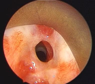
Subglottic stenosis (( can mistaken with asthma!!))

Partial or complete narrowing of the subglottic area may be congenital or acquired. The problem is rare and challenging, affecting soft tissue and cartilage support.
Iatrogenic injuries cause most of the problems seen. Often, subglottic stenosis has an insidious onset, and early manifestations are usually mistaken for other disorders (eg, asthma, bronchitis).
An image depicting subglottic stenosis can be seen below.

Preoperative view of subglottic stenosis via an endoscopic approach.
Preoperative view of subglottic stenosis via an enPreoperative view of subglottic stenosis via an endoscopic approach. 

Pathophysiology:
Congenital stenosis has two main types, membranous and cartilaginous.
In membranous stenosis, fibrous soft tissue thickening is caused by increased connective tissue or hyperplastic dilated mucus glands with absence of inflammation. 
Membranous stenosis is usually circumferential and may extend upward to include the true vocal folds.
In cartilaginous stenosis, a thickening or deformity of the cricoid cartilage most commonly occurs, causing a shelflike plate of cartilage and leaving a small posterior opening. 
Cartilaginous stenosis is less common than membranous stenosis.
Acquired subglottic stenosis is secondary to localized trauma to subglottic structures. Usually, injury is caused by endotracheal intubation or high tracheostomy tube placement. If irritation persists, the original edema and inflammation progress to ulceration and granulation tissue formation. This may or may not involve chondritis with destruction of the underlying cricoid cartilage and loss of framework support.
When the source of irritation is removed, healing occurs with fibroblast proliferation, scar formation, and contracture, leading to stenosis or complete occlusion of the airway.

Presentation:
Adults with mild congenital stenosis are usually asymptomatic, and they are diagnosed after a difficult intubation or while undergoing endoscopy for other reasons.
Patients with acquired stenosis are diagnosed from a few days to 10 years or more following initial injury. The majority of cases are diagnosed within a year. Symptoms include the following:

* Dyspnea (may be on exertion or with rest, depending on severity of stenosis)
* Stridor
* Hoarseness
* Brassy cough
* Recurrent pneumonitis
* Cyanosis
Many patients would have been diagnosed with asthma and recurrent bronchitis prior to discovery of stenosis. A high index of suspicion is warranted with the onset of respiratory symptoms following intubation, regardless of the duration of intubation.

Source:medscape

Friday, 16 September 2016

Alzheimer disease (AD)


.Alzheimer's disease (AD) is the most common form of dementia among older people.


 Dementia is a brain disorder that seriously affects a person's ability to carry out daily activities.

AD begins slowly.


 It first involves the parts of the brain that control thought, memory and language. 


People with AD may have trouble remembering things that happened recently or names of people they know. A related problem, mild cognitive impairment (MCI), causes more memory problems than normal for people of the same age. Many, but not all, people with MCI will develop AD.

In AD, over time, symptoms get worse. People may not recognize family members. They may have trouble speaking, reading or writing. They may forget how to brush their teeth or comb their hair. 


Later on, they may become anxious or aggressive, or wander away from home. Eventually, they need total care. This can cause great stress for family members who must care for them.

AD usually begins after age 60. The risk goes up as you get older. Your risk is also higher if a family member has had the disease.

No treatment can stop the disease. However, some drugs may help keep symptoms from getting worse for a limited time.

Thursday, 15 September 2016

Dermatographia - skin writing !!-


Dermatographia is a condition also known as skin writing.

 When people who have dermatographia lightly scratch their skin, the scratches redden into a raised wheal similar to hives.
These marks usually disappear within 30 minutes.


The cause of dermatographia is unknown, but it can be triggered in some people by infections, emotional upset or medications such as penicillin.

Most people who have dermatographia don't seek treatment.

 If your signs and symptoms are especially bothersome, your doctor may recommend allergy medications such as cetirizine (Zyrtec) or diphenhydramine (Benadryl).

.
www.mayoclinic.org

Tongue cancer

Tongue cancer

Tongue cancer is a type of oral cancer that forms in the front two-thirds of the tongue. Cancer that forms in the back one third of the tongue is considered a type of head and neck cancer.

Tongue cancer usually develops in the squamous cells, the thin, flat cells that cover the surface of the tongue.

Common tongue cancer symptoms

Symptoms of tongue cancer are very similar to symptoms of other types of oral cancer. It can often be mistaken for a cold that won’t go away, or a persistent sore in the mouth. Other tongue cancer symptoms and signs may include:

Persistent tongue and/or jaw pain
A lump or thickening in the inside of the mouth
A white or red patch on the gums, tongue, tonsil, or lining of the mouth
A sore throat or feeling that something is caught in the throat that does not go away
Difficulty swallowing or chewing
Difficulty moving the jaw or tongue
NOTE: These symptoms may be attributed to a number of conditions other than cancer. It is important to consult with a medical professional for an accurate diagnosis.

Advanced treatments for tongue cancer

Common tongue cancer treatments include:

Surgery: Tumor resection involves an operation to remove the entire tumor from the tongue. Minimally invasive surgical techniques are used whenever possible to treat tongue cancer.

Radiation therapy: Your radiation oncologist will administer radiation therapy to cancerous tissues of the tongue, using a high dose with pinpoint accuracy, sparing healthy tissue and shortening procedure times.

Chemotherapy: Often combined with radiation therapy, chemotherapy uses anticancer drugs to destroy cancer cells throughout the body. It may be an option if the cancer has spread to nearby lymph nodes. Different chemotherapy drugs can be combined to attack cancer cells at varying stages of their growth cycles and decrease the chance of drug resistance.

Targeted drug therapy: Targeted drug therapy targets cancerous cells to interfere with cell growth on a molecular level. It is often combined with chemotherapy and/or radiation therapy as part of a tongue cancer treatment plan.

Copied

Wednesday, 14 September 2016

Myocardial infarction


Myocardial infarction (MI) (ie, heart attack) is the irreversible death (necrosis) of heart muscle secondary to prolonged lack of oxygen supply (ischemia).


Overview

Practice Essentials

Myocardial infarction (MI) (ie, heart attack) is the irreversible death (necrosis) of heart muscle secondary to prolonged lack of oxygen supply (ischemia).

Acute myocardial infarction, reperfusion type. In this case, the infarct is diffusely hemorrhagic. There is a rupture track through the center of this posterior left ventricular transmural infarct.

The mechanism of death was hemopericardium.

Signs and symptoms

Patients with typical MI may have the following prodromal symptoms in the days preceding the event (although typical STEMI may occur suddenly, without warning):
*Fatigue


*Chest discomfort


*Malaise


Typical chest pain in acute MI has the following characteristics:
*

Intense and unremitting for 30-60 minutes

Retrosternal and often radiates up to the neck, shoulder, and jaw and down to the ulnar aspect of the left arm

Usually described as a substernal pressure sensation that also may be characterized as squeezing,

* aching, burning, or even sharp.


In some patients, the symptom is epigastric, with a feeling of indigestion or of fullness and gas


The patient’s vital signs may demonstrate the following in MI:


The patient’s heart rate is often increased secondary to a high sympathoadrenal discharge.


The pulse may be irregular because of ventricular ectopy, an accelerated idioventricular rhythm, ventricular tachycardia, atrial fibrillation or flutter, or other supraventricular arrhythmias; bradyarrhythmias may be present

In general, the patient's blood pressure is initially elevated because of peripheral arterial vasoconstriction resulting from an adrenergic response to pain and ventricular dysfunction.

http://emedicine.medscape.com


Cell envelope!


The cell envelope is a protective layer of armor that surrounds the bacterium and allows it to survive in diverse and extreme environments.

The cell envelopes of some bacteria consist of a cytoplasmic membrane surrounded by a tough and rigid mesh called a cell wall ; these bacteria are referred to as gram-positive bacteria.
 In contrast, the cell envelope of a gram-negative bacterium consists of a cytoplasmic membrane surrounded by a thin cell wall that is itself surrounded by a second lipid membrane called the outer membrane.
 The outer membrane contains large amounts of lipopolysaccharide (LPS), a molecule that is very toxic to humans. The space between the outer membrane and the cytoplasmic membrane, which contains the cell wall, is called the periplasmic space or the periplasm.

Whether a bacterium is gram- positive or gram-negative can usually be determined by a technique called Gram stain- ing, which colors gram-positive bacteria blue or purple and gram-negative bacteria pink.
 Gram staining is often the first step used by a hospital microbiology laboratory in identifying an unknown bacterium from a clinical specimen.

As in human cells, the cytoplasmic membrane prevents ions from flowing into or out of the cell itself and maintains the cytoplasm and bacterial components in a de- fined space.
 The cell wall is a tough layer that gives a bacterium its characteristic shape and protects it from mechanical and osmotic stresses.

 In gram-negative bacteria, the outer membrane acts as an additional protective barrier and prevents many substances from penetrating into the bacterium. This layer, however, does contain channels called porins that allow some compounds such as molecules used in metabolism by the bac- terium to pass through.

Since human cells do not possess a cell wall, this structure is an ideal target for antimicrobial agents. To appreciate how these agents work, we must first understand the structure of the cell wall. This complex assembly is made up of a substance called peptidoglycan, which itself consists of long sugar polymers. The polymers are repeats of two sugars: N-acetylglucosamine and N-acetylmuramic acid
. If the cell wall were to consist of these polymers alone, it would be quite weak.

 However, peptide side chains extend from the sugars in the polymers and form cross-links, one peptide to another. These cross-links greatly strengthen the cell wall, just as cross-linking of metal loops strengthened the chain mail armor used by medieval knights. 

The cross-linking of peptidoglycan is mediated by bacterial enzymes called penicillin-binding proteins (PBPs). (The reason for this nomenclature will become apparent in later chapters.) These enzymes recognize the terminal two amino acids of the peptide side chains, which are usually d-alanine–d-alanine, and either directly cross-link them to a second peptide side chain or indirectly cross-link them by forming a bridge of glycine residues between the two peptide side chains.

The formation of a tough cross-linked cell wall allows bacteria to maintain a char- acteristic shape. For example, some bacteria are rod shaped and referred to as bacilli. Cocci are spherical in shape. Coccobacilli have a morphology that is intermediate between that of bacilli and cocci. Finally, spirochetes have a corkscrew shape.